The Layers of a Dog Eye:
The dog eye has three different layers:
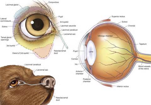
- Fibrous Tunic: This part forms the outer most opaque layer and is comprised of collagen and fibrous tissues, called sclera. Sclera majorly forms the posterior of the eye, while anterior or the front part of the eye is the cornea, which is transparent and allows entry of light into the eye.
- Vascular Tunic: The dog eye is supplied with blood vessels, which supplies oxygen and nutrients to the all parts of the eye through the blood. The major part of the vascular tunic comprises a network of blood vessels, and also the choroid, which lies beneath the sclera, which helps in adjustment of the lens and “accommodation" or muscular constriction and relaxation which controls light. It is believed that this muscular mechanism is influenced by the vascular supply in the eye.
- Nervous Tunic: This functional part of the eye is controlled and is related to the nervous system of the dog eye anatomy. Photoreceptor cells called “retina" lies on the whole round wall of the posterior eye. These are controlled by retina, which collects and identifies the light, which is then processed to the brain with help of the “optic nerve". The “Blind spot" or “Optic Disc" is the dark spot on the retinal wall, which has no photoreceptor cells over it and this part opens into the optic nerve duct, which contains a rich nerve supply, that connects the eye with the brain.

The functionality of the eye enhanced with various anatomical features, which includes the, eyelids, eyelashes, conjunctiva, cornea, iris, pupil, lens, third eyelid (nictitans gland) lacrimal gland and viteous chamber of the eye.
The remaining parts are:
- Eyelids and Eyelashes: These are the outer most protective layers of the dog eye anatomy, which are flexible and wipe the tears over the eye surface to keep it protected and away from foreign particles such as dust and debris. The eye blinks as an involuntary action, which is controlled by the brain. These parts also help to control light rays entering the eye.
- Cornea: The cornea is the central dome shaped area of the eye surface, which is supposed to be a pathway for light entering into the eye. The cornea not only functions as a protective part of the inner eye but also it focuses light on the retina at the back of the eye. The focusing and light control function of the cornea is facilitated by the iris.
- Conjunctiva: Tough outer protective layers of the eye, the sclera is covered by a thin membrane, called conjunctiva. This thin membrane is located near the front of the eye. Conjunctiva runs over the cornea and covers all of the inner parts of the eyelids.
- Iris: This is the circular colored area of the eye, which controls the amount of light entering into the eye. Due to the functionality of this eye part, the pupil gets smaller or larger. The pupil is the central black area of the eye, which dilates or constricts under the influence of the iris and environment, to let more or less light entering into eye. The pupil dilates in a darker environment and constricts in a bright environment to control and allow an adequate amount of light to enter the eye.
- Lens: The lens is just behind the iris of the eye, which is meant for focusing nearby or distant objects on the retina. The function of the lens is controlled by the ciliary muscles which contract, causing the lens to become thicker and nearby objects more focused. On the other hand, if the ciliary muscles relax, the lens becomes thinner and thus distant objects are focused on the retina.
- Retina: These are photoreceptor cells, which are distributed over the round back surface wall of the eye. The most dense part of the photoreceptor tissues or retina of the dog eye are called area centralis. In this region, thousands of photoreceptor cells are packed making the incoming images sharper. Each retina photoreceptor cell is attached with a nerve, which enters into the optic duct, through the optic disc and is connected to the brain. All the minor optic nerves of the retina are bundled into a larger optic nerve. Retinal tissues convert images into electrical impulses, which are carried to the brain with the help of the optic nerves.
- Nictitaing Membrane: In dogs, the eye is not only protected by the eyelids, but also with a nictitating membrane, which is also called the third eyelid. This is whitish to pink in color and is located on the edge of the inner part of the eyelids, near the nose. This membrane extends to the surface of the eye when the protection is needed or when the eye ball becomes vulnerable to scratches, irritation and in case of inflammation.
- Lacrimal Glands: These are tear producing glands. Tears keep the dog eyes wet and lubricated. Debris, dust etc is flushed out of the eye with the help of these tears. There are two types of Lacrimal glands, of which the lacrimal glands produce watery content or tears and the mucous glands in the conjunctiva produce mucous which mixes with tears to forms the protective nature of tears, as antibacterial or the slippery nature of tears wipe the debris out of the eye. The mucous part of the tears helps in preventing them from rapid evaporation. Tears are drained with the help of nasolacrimal ducts, which open into the nose.
Eye Conditions with links to more in-depth articles
Allergies
Itchy, red, swollen, tearing eyes may mean eye allergies. Also see today's AccuWeather pollen count map.
Amblyopia (Lazy Eye)
Amblyopia (or "lazy eye") is a vision development problem in infants and young puppies that can lead to permanent vision loss.
Bell's Palsy in Dogs
This condition causes sudden paralysis of one side of the face.
Blepharitis
Inflammation of the eyelids can cause chronic eye irritation, tearing, foreign body sensation and crusty debris.
Cataracts
If you live long enough, it's likely you eventually will notice cloudy vision from cataracts.
Chalazion
A swollen bump in the eyelid could be a chalazion. Learn about causes and treatments.
Color Blindness
Dog's aren't really color blind, it's really more of a lack of brightness and range.
Corneal Ulcer
A corneal ulcer is a wound on the surface (called the epithelium) of your dog's eye and possibly some of the surrounding tissue (stroma).
Detached Retina
If both retinas are detached, it is most likely a sign of a more serious underlying medical condition. Glaucoma, for instance, is one such condition. Exposure to certain toxins can also cause the retina to detach.
Dry Eye Syndrome
Dry eye (keratoconjunctivitis sicca or KCS) is a painful and potentially dangerous condition caused by the inadequate production of tears.
Eye Occlusions (Nasolacrimal occlusion in dogs)
Blockage of a tear duct.
Nystagmus and Eye Weeping in Dogs
Uncontrollable eye movements from nystagmus often have neurological causes.
Ocular Rosacea
How to solve eye and eyelid irritation.
Optic Neuritis and Optic Neuropathy
An inflamed optic nerve can cause blurry vision and blind spots.
Photophobia (Light Sensitivity)
Sensitive to light? Cone degeneration is one of the many causes in dogs.
Pinguecula and Pterygium
Pingueculae and pterygia are growths on the eye.
Ptosis (Drooping Eyelid or Entroption)
Drooping eyelids or the rolling in or out of the eyelids.
Strabismus & Cross eyes
Key facts about strabismus and crossed eyes, including causes and treatments (strabismus surgery, vision therapy and orthoptics).
Subconjunctival Hemorrhage or Red Eye
Sudden redness in the white of your eye may be a subconjunctival hemorrhage.
Eye Diseases with links to more in-depth articles
Conjunctivitis (Pink Eye)
What you can do about redness, swelling, itching and tearing of pink eye. Also read about pink eye treatment and the various conjunctivitis types.
Diabetic Retinopathy
Diabetes causes sight-threatening retinal degradation.
Corneal Dystrophy
Causes loss of vision and clouding of the cornea due to degeneration of the corneal endothelium and corneal edema.
Glaucoma
Glaucoma damages the optic nerve and diminishes the field of vision.
Macular Degeneration (PRA)
There are multiple forms of PRA which differ in the age of onset and rate of progression of the disease. Some breeds experience an earlier onset than others; other breeds do not develop PRA until later in life.
Macular Dystrophy
Central vision loss can be associated with this inherited eye disease.
Retinitis Pigmentosa
Poor night vision and a narrowing field of vision.
Surgical Eye Procedures with links to more in-depth articles
Cataract Surgery
It's the most common eye surgery for pets in the United States.
Cornea Transplant
First corneal transplant on a dog in the US.
Glaucoma Surgery
Glaucoma surgery for pets.
Vitrectomy & Vitreoretinal Procedures
These delicate surgical procedures for macular holes, retinal detachments and other conditions are performed in the eye's deep interior.


